
History
This is a 58 year old woman who presents with a crusting pigmented lesion of the nipple of the left breast. The clinical impression was a melanoma.
Microscopic Findings
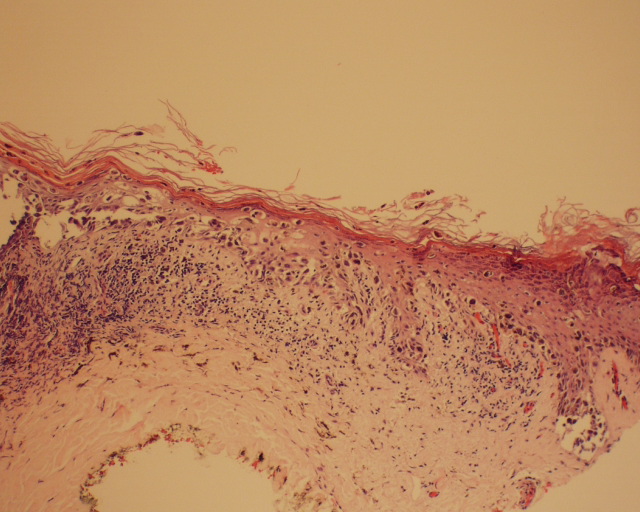 |
Sections show an atypical intraepidermal proliferation of both individual and nested cells, many of which contain abundant cytoplasmic melanin pigment. | |||
| Figure 1 |
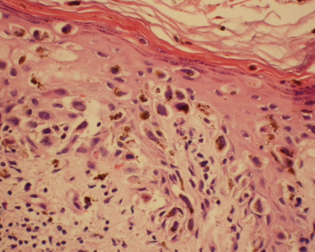 |
Pigment incontinence is noted within the stratum corneum as well. The dermal-epidermal junction shows discohesive clefting with atypical cells associated with melanin pigment. No definite glandular differentiation is noted. | |||
| Figure 2 |
Immunoperoxidase and Special Stains
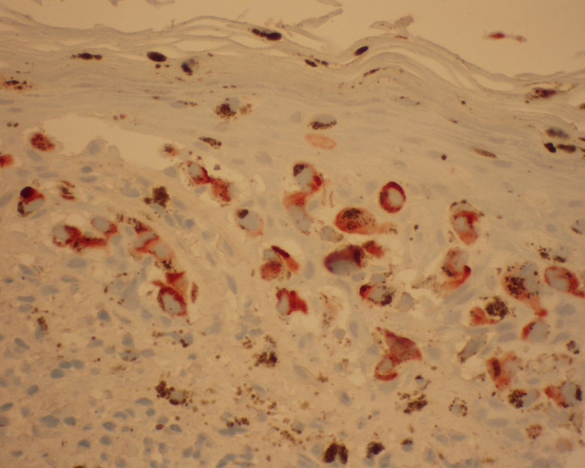 |
Immunoperoxidase studies showed a positive EMA within the atypical cells. | |||
| Figure 3 |
What is your diagnosis?
Last Updated 11/2/2002
Send mail to The Doctor's Doctor with questions or comments about this web site.
Copyright © 2004 The Doctor's Doctor